Türkçe
A splint is a medical device used to immobilize and support a part of the body that has been injured or undergone a surgical procedure. Its primary function is to support the healing process by keeping damaged tissues such as bone, cartilage, or joints immobile in the correct position. In this way, it both reduces pain in the area and protects sensitive tissues against external factors, creating an ideal environment for healthier and faster repair. It plays an indispensable role, especially in trauma such as fractures and sprains and in postoperative care, by enabling tissues to unite and heal in the correct anatomical position.
What Is a Splint?
A splint is a medical support device used to immobilize and protect a joint and support healing in cases of bone fractures, dislocations, sprains, or soft tissue injuries. It is more flexible and removable than a cast and is generally made of plastic, metal, or fabric materials. Splints are preferred in the early period after trauma to reduce swelling and limit joint movement.
Why Is a Splint Used and What Does It Do?
When we see someone with their arm or leg wrapped after an accident or sports injury in daily life, the first word that comes to mind is “cast.” However, in medicine, a cast is not the only method used to immobilize an area. This is where the “splint,” which is often confused with a cast but has very different and critical functions, comes into play. In the most basic sense, a splint is a medical device designed to protect and support a damaged part of the body and help it heal by keeping it immobile in the correct position. Its purpose is both to accelerate the healing process and ensure that it is completed in the healthiest possible way.
What Is the Difference Between a Splint That Immobilizes the Body and a Cast?
The most fundamental and important feature that distinguishes a splint from a cast is its structure. A cast completely surrounds a limb like a cylinder, while a splint is generally formed by securing a rigid support plate to the area with elastic bandages or hook-and-loop straps. This allows part of the splinted area to remain exposed so that the skin can breathe. This structural difference, which may seem simple, is actually the basic principle that determines the purpose and importance of a splint.
Especially immediately after an injury, during the acute period, the body’s first response is to develop significant swelling, or edema, in that area. This is a natural part of the healing process. Now imagine wrapping an area with the potential to swell in a completely enclosed and rigid cast. As the swelling increases, the tissues inside the cast begin to be compressed as if in a vise. This may cause the blood vessels carrying blood to the tissues and the nerves providing sensation to be crushed. In the worst-case scenario, a very dangerous condition called “compartment syndrome” may develop, in which tissue nutrition is completely impaired and emergency surgical intervention is required, potentially even leading to loss of the limb.
This is where the brilliance of the splint becomes apparent. Since it is not completely enclosed, it allows space for this natural tissue swelling. In this way, it provides the necessary immobilization while eliminating serious risks related to compression. Therefore, in emergency departments or immediately after surgery, during the initial period when swelling is expected to be most intense, the preferred method is generally a splint. Splints may be available as ready-made, prefabricated devices for common injuries or may be shaped by the physician according to the patient’s anatomy. In both cases, the aim is to preserve the correct anatomical position of the injured area and create the most ideal conditions for the body’s own repair mechanisms.
What Are the Basic Functions of Splints and In Which Situations Are They Used?
There are several basic therapeutic goals behind the application of a splint. These goals are highly important at every stage of the healing process.
The main functions of a splint are as follows:
- Restricting movement (Immobilization)
- Reducing pain
- Protecting soft tissues
- Providing an ideal healing environment
- Compensating for muscle weakness
Thanks to these functions, splints have a very wide range of uses. They play a life-saving role not only in fractures but also in many different conditions.
Some common situations in which splints are used.
- Fractures
- Sprains
- Muscle and tendon strains
- Joint dislocations
- Serious soft tissue injuries
- Deep cuts crossing a joint
- Suspected hidden (occult) fractures
- Arthritis flare-ups
Tendinitis and tenosynovitis (inflammation of the tendon and its sheath)
Which Materials Are Used to Make Splints?
Advances in medical technology have also diversified the materials used in splint production. Immobilization procedures that were once performed with primitive materials such as wood and bamboo are now carried out using much more functional and comfortable materials. Each material has its own advantages and areas of use:
- Plaster: It is the most traditional material. It consists of bandages impregnated with plaster powder that hardens when combined with water. Its greatest advantage is that it can be shaped very easily while wet, allowing the physician to create a mold that fits the contours of the patient’s body exactly. However, it is heavy, deteriorates easily when wet, and may partially obstruct the image on X-rays.
- Fiberglass: It is a more modern alternative to plaster. It is made of fiberglass fabric impregnated with a water-activated resin. It is much lighter, more durable, and stronger than plaster. Its porous structure allows the skin to breathe, and it is more water-resistant. It is also more radiolucent than plaster. Its only disadvantage is that it hardens more quickly, leaving less time for shaping.
- Thermoplastics: These materials, used especially in sensitive areas such as the face and hand, have revolutionized aesthetic and functional splinting. These normally rigid plastic sheets become soft and easily moldable like dough when heated in hot water, generally at 60-70°C. This allows a personalized mold to be created directly on the patient’s nose, hand, or face. When cooled, they harden quickly and become an extremely lightweight and durable splint. They are among the most frequently preferred materials for external nasal splints.
- Silicone: It is a material that is fully compatible with the body, biocompatible, flexible, and chemically nonreactive. These characteristics make it ideal, especially for splints placed inside the body. Internal nasal splints used after nasal surgery are generally made of silicone. Thanks to its soft and non-stick surface, it does not damage the nasal mucosa and is much easier to remove.
- Aluminum: Easily bendable aluminum plates supported with pads are used as a practical solution, especially in ready-made, prefabricated splints such as finger or external nasal splints.
Why Are Both Internal and External Splints Used in Nasal Surgery?
The nose is an organ located in the center of the face and has great importance both aesthetically and functionally. Therefore, the healing process after nasal surgery is at least as critical to the success of the result as the surgery itself. In procedures such as rhinoplasty, aesthetic nose surgery, and septoplasty, surgery for internal nasal deviation, an approach generally called “dual splinting” is adopted. This refers to the combined use of internal and external splints, each serving a different purpose.
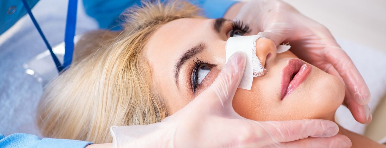
As the name suggests, the external nasal splint is a protective mold placed on the external surface of the nose over the skin. Its primary function is to protect the nasal bones and cartilage framework reshaped by surgery against external impacts like a shield and keep them stable in their new positions.
The internal nasal splint, or septal splint, consists of thin, flexible plates placed through the nostrils on both sides of the septum, which is the central partition of the nose. Its function is entirely inside the nose: to support the healing septum, prevent blood accumulation, or hematoma, and most importantly, prevent intranasal adhesions, or synechiae. These two splints support the nose both internally and externally, enabling it to heal in the most ideal way.
What Does an External Nasal Splint Do?
The small mold, usually skin-colored, placed over the nose after rhinoplasty, namely the external splint, plays a key role in preserving the aesthetic result. It is not merely a simple protector; it has several important functions:
- The primary functions of the external nasal splint.
- Stabilizing the new shape
- Protecting against external impacts
- Helping reduce swelling and bruising
- Helping the skin adapt to the new framework
- Preventing involuntary touching
Its application generally involves first applying thin sterile tapes to the nasal skin, then placing a heat-molded thermoplastic or manually shaped aluminum splint and securing it with additional tapes. This splint generally remains in place for 7 to 10 days after surgery and is removed by the physician during a clinical follow-up. This period is sufficient for the bones and cartilage to begin their initial healing.
What Are the Vital Benefits of Internal Nasal Splints?
Internal nasal splints used after surgeries involving the nasal septum, such as septoplasty or septorhinoplasty, provide critical benefits that the patient may not notice but that directly affect the result. These thin silicone plates play a major role in ensuring a smooth postoperative period.
The most important benefits provided by internal nasal splints are as follows:
- Stabilizing the septum in the midline
- Preventing septal hematoma, or blood accumulation
- Preventing intranasal adhesions, or synechiae
- Helping control bleeding
Perhaps the most important and scientifically best-proven benefit is preventing the formation of synechiae, or adhesions. After surgery, the mucosal surfaces of the septum and the opposing nasal turbinate are injured. If these two injured surfaces come into contact while healing, scar tissue may form between them like a permanent bridge, causing lifelong nasal obstruction. The internal splint places a physical barrier between these two surfaces and definitively prevents them from adhering. Many modern silicone splints used today also contain air channels that allow the patient to breathe through the nose after surgery. Compared with old-style nasal packing, this innovation greatly increases patient comfort. These splints are generally removed after 3 to 7 days, again in the clinic, with a simple procedure.
Is the Use of an External Splint Necessary After Every Rhinoplasty?
In medicine, absolute rules such as “always” or “never” rarely apply, and this has also begun to be true for the use of an external splint after rhinoplasty. This practice, which was accepted as a standard for many years, has been questioned in recent years in light of scientific studies. Traditionally, it was believed that the external splint kept the bones in place and reduced swelling. However, it has been observed that strong scientific evidence supporting this widespread belief is actually quite limited.
Recent comprehensive studies and systematic reviews have not found clear evidence that the use of an external splint significantly reduces postoperative bruising and swelling. In fact, some studies have observed that bones successfully healed in the desired position even in patients who received only special taping techniques and no splint at all. This suggests that the necessity of the splint may have been overstated and that meticulous surgical technique may be sufficient in many cases.
In addition, the potential disadvantages of external splint use should not be ignored:
Possible risks of the external splint:
- Pressure-related skin wounds
- Skin necrosis (tissue death)
- Contact dermatitis (contact eczema)
- Unwanted depressions on the nasal bridge
Can Ear Deformities in Babies Be Corrected With a Non-Surgical Splint?
Yes, the answer to this question is definitely yes, and this is one of the most exciting and least-known applications among aesthetic approaches. The first few weeks and months of life provide a magical “window of opportunity” during which ear cartilage is soft and moldable almost like modeling clay. The reason for this is the high level of maternal estrogen hormone that remains in the baby’s circulation after birth. This hormone temporarily makes the cartilage extremely flexible.
In this method, called neonatal ear molding, specially designed small and soft splints, or molds, are placed on the baby’s ear. These splints apply gentle but continuous pressure to the cartilage, guiding it toward the ideal anatomical shape it should have. Over time, as the maternal estrogen levels decrease and the baby’s cartilage naturally begins to harden, the new shape given by the splint becomes permanent.
- Some congenital ear deformities that can be corrected with this method.
- Prominent ear
- Stahl’s ear, a pointed deformity also known as “Spock ear”
- Cryptotia, where the upper part of the ear is buried beneath the skin
- Helical fold abnormalities
- Lop ear
This approach is extremely successful when applied early, ideally within the first 1-2 months. Being painless, safe, and most importantly non-surgical makes it an invaluable alternative that can spare the child from the risks, cost, and need for general anesthesia associated with future prominent ear surgery, or otoplasty.
What Is the Role of Bandages and Headbands After Prominent Ear Surgery?
After prominent ear surgery performed on older children or adults, the principles of splinting come into play to ensure the permanence of the surgical result. This process consists of two stages. Immediately after surgery, a bulky and compressive dressing that completely covers the ears, also called a “turban dressing,” is applied around the patient’s head. The purpose of this initial dressing is to prevent blood accumulation, or hematoma, behind the ear, keep swelling under control, and most importantly, protect the sensitive ears from any impact or bending during the first critical days.
This initial dressing is generally removed in the clinic after several days to one week. The patient then moves on to the second and longer-term splinting stage: an elastic headband. Patients are asked to wear this headband, which resembles an athletic band, continuously during the first week and then only while sleeping at night for the following several weeks. The function of this headband is to act as a night splint, especially during sleep, preventing the person from unknowingly rubbing the ears against the pillow, bending them, or folding them forward.
The logic behind this is a biomechanical phenomenon called “cartilage memory.” Mature ear cartilage, much like a piece of plastic that has been bent and released, tends to return to its former, “remembered” prominent position even after being surgically reshaped and secured with sutures. The postoperative headband applies a gentle external force that continuously counteracts this tendency to return. In this way, the cartilage heals in its new position, strong scar tissue forms around the sutures, and the aesthetic result becomes permanent.
What Are the Other Splint Applications in the Jaw and Facial Region?
The use of splints is not limited to the nose and ears. They also play vital roles in fields such as maxillofacial surgery and dentistry, which concern the functional and aesthetic integrity of the face.
- Orthognathic Surgery (Jaw Surgery): In these major operations aimed at changing the position of the upper and lower jaws, acrylic splints called “occlusal splints” or “wafers” are used. These splints are not merely supports but serve as a roadmap for the surgery itself. During the operation, the surgeon positions and fixes the jawbones that have been cut and moved so that they fit precisely into this pre-prepared splint. The splint ensures the ideal occlusion of the teeth and guarantees that the surgical plan is translated into reality.
- Temporomandibular Joint Disorders (TMD): Intraoral splints, generally known as night guards, are frequently used in the treatment of problems such as pain, clicking sounds, and difficulty chewing in the jaw joint. These splints reduce involuntary muscle activity such as teeth clenching, or bruxism, thereby relieving excessive stress on the jaw joint and chewing muscles. In this way, they both protect the teeth from wear and allow the joint to rest and relax.
- Obstructive Sleep Apnea (OSA): In patients with snoring and pauses in breathing during sleep, a Mandibular Advancement Splint, also known as a snoring appliance, is an effective treatment option. This device is a splint with a special mechanism that fits over the upper and lower teeth and moves the lower jaw forward by several millimeters in a controlled manner during sleep. By bringing the lower jaw forward, it also moves the base of the tongue and soft palate forward. This movement widens the airway in the throat and prevents it from collapsing during sleep, thereby eliminating both snoring and pauses in breathing.
What Should a Patient With a Nasal Splint Pay Attention To?
The postoperative period is at least as important as the surgery itself, and the greatest responsibility during this process falls on the patient. Following the rules below accelerates the healing process, reduces the risk of complications, and ensures the best possible result.
Position and Activity Rules
- During the first week, sleep with your head elevated at an angle of 30-45 degrees using 2-3 pillows.
- Do not bend forward, strain, or lift anything heavy, more than 3-4 kg.
- Keep your mouth open when sneezing to reduce pressure.
- Do not blow your nose until your doctor permits it, generally for 2-3 weeks.
Hygiene and Care
- Make every effort not to get the external splint and tapes wet.
- Gently clean the nostrils and any suture lines 3-4 times a day as recommended by your doctor, generally with a cotton swab containing hydrogen peroxide followed by antibiotic ointment.
- Use the saline sprays recommended by your doctor regularly to keep the inside of your nose moist and soften crusts.
Situations in Which You Should Contact Your Doctor Immediately
- Fever above 38.5°C
- Severe and throbbing pain that cannot be controlled with medication
- Uncontrollable, bright red fresh bleeding
- Foul-smelling, yellow-green discharge from the nose
- Displacement or detachment of the splint

Prof. Dr. Murat Songu – Rhinoplasty (Nose Aesthetics) Specialist
Prof. Dr. Murat Songu was born in 1976 in Izmir and completed his medical education at the Ege University Faculty of Medicine. He then completed his residency training in the Department of Otorhinolaryngology at Celal Bayar University. Between 2005 and 2006, he received advanced training in rhinoplasty, functional nasal surgery, and skull base surgery in Bordeaux, France, working with leading rhinologists such as Prof. Vincent Darrouzet and Dr. Guy Lacher.
Prioritizing natural appearance, the preservation of breathing function, and facial aesthetic harmony, Prof. Dr. Songu is nationally and internationally recognized for his expertise in open rhinoplasty, piezo (ultrasonic) rhinoplasty, revision rhinoplasty, tipplasty, and functional septorhinoplasty. He has participated as an instructor and speaker at numerous rhinoplasty congresses worldwide.
With over 100 scientific publications, book chapters, and more than 1700 citations, Prof. Dr. Murat Songu is one of Türkiye’s most respected academics in the field of rhinoplasty, combining natural, facially harmonious, and functional results with scientific and aesthetic excellence.