Türkçe
Cartilage, the flexible yet durable structural tissue of our body, is a specialized connective tissue that is not as hard as bone but is stronger than muscle. Its primary functions are to create a smooth surface in joints so that bones can move without friction, act as a cushion by absorbing impacts, and provide structural support and shape to organs such as the nose and ears. To perform these versatile functions, there are three main types in the body, each with different mechanical properties: hyaline, elastic, and fibrocartilage. The unique structure of this tissue gives it both strength and flexibility.
What Is Cartilage?
Cartilage is a type of flexible yet durable connective tissue found at the ends of bones, in joints, the ears, nose, and respiratory tract. It is softer than bone, has no blood vessels, and receives nourishment through diffusion from surrounding tissues. Articular cartilage reduces friction between bones and increases mobility by absorbing impacts. When damaged, it is difficult to heal.
What Is Cartilage, This Flexible Structural Tissue of Our Body?
We can think of cartilage as a unique structural material in the body that is neither as hard as bone nor as soft as muscle. It would not be wrong to compare it to durable rubber that does not lose its flexibility. Thanks to this unique structure, it can perform many tasks that may seem contradictory at the same time.
Its best-known function is to cover the surfaces of bones facing each other in the joints like a smooth cushion. Just like the perfectly lubricated gears of a machine, our bones move smoothly, painlessly, and without rubbing against one another thanks to cartilage. It absorbs the pressure and shock generated during actions such as walking, running, or jumping like a car’s suspension system, protecting the underlying bone tissue from the damaging effects of impact.
However, the capabilities of cartilage are not limited to this. In areas where both flexibility and structural integrity are required, it acts almost like a framework. For example, the shape of our nose is largely due not to the underlying bone but to its cartilage framework. This framework gives the nose a certain degree of flexibility against impacts while also providing its characteristic form. Similarly, the complex and aesthetic curves of the auricle are possible entirely because of elastic cartilage. In the trachea, rings of cartilage arranged one above another ensure that this vital airway never collapses, allowing us to breathe.
From a developmental perspective, its role is even greater. While we are in the womb, a large part of our skeleton is actually made of cartilage, which serves as a preliminary model or “blueprint” for bone. Over time, this cartilage model is gradually replaced by bone tissue. Traces of this remarkable transformation continue to exist in adulthood on our joint surfaces and in the growth plates that allow our bones to lengthen.
What Is Inside Cartilage and How Does This Structure Make It So Special?
The secret of cartilage’s impressive mechanical properties lies in its structure. We can imagine cartilage as a structure consisting of a very small number of “worker cells” living inside a massive gel-like house that they themselves produce.
The worker cells inside this house are specialized cartilage cells called chondrocytes. Although they make up only a small portion of the tissue’s total volume, they are responsible for all maintenance and repair of cartilage. We can think of them as tiny self-sufficient factories that continuously renew and repair the structure around them. These cells are highly sensitive; they can detect even the slightest mechanical or chemical change in their surroundings and adjust their production accordingly. This “intelligent” behavior is vital for maintaining the health and integrity of cartilage tissue. When we perform cartilage transplantation in surgery, we must never forget that we are actually moving these living factories to a new location and that preserving their health is essential for success.
Why Can Cartilage Not Heal Itself When It Is Damaged?
One of the most interesting and clinically important characteristics of cartilage is its “isolation.” Unlike other tissues in the body, cartilage lacks three important structures: blood vessels, nerve fibers, and lymphatic vessels. This has both advantages and disadvantages:
The greatest disadvantage of this isolation is that cartilage has almost no ability to repair itself. When an area of the body is cut, blood vessels immediately begin a healing process by carrying repair cells, nutrients, and oxygen to the region. Cartilage does not have this “logistical support network.” It receives its nutrients through very slow seepage, or diffusion, from surrounding tissues. Therefore, once it is damaged, it has no mechanism to repair that damage, and the damage is generally permanent. Since it contains no nerve fibers, cartilage damage itself does not directly cause pain. The pain felt in cartilage wear in the joints generally results from involvement of the underlying bone or inflammation of other surrounding tissues.
However, there is another side to the coin. While this isolation makes cartilage a “problematic tissue” in terms of healing, it also makes it a “perfect guest” for surgical repairs. Cartilage that we take from one part of the body and transplant, or graft, to another during surgery is largely hidden from the body’s immune system because it contains no blood vessels. Since immune cells move through the bloodstream, they cannot easily detect and attack this new tissue. This makes cartilage “immunologically privileged” and reduces the risk of rejection of cartilage grafts, especially those taken from the patient’s own body, to nearly zero. In its new location, the graft continues to survive on nutrients diffusing from surrounding tissues, just as it did in its original location. Thanks to this paradox, we can create highly safe, long-lasting, and stable structures using cartilage grafts.
What Types of Cartilage Are Found in Our Body?
Not all cartilage in our body is the same. Just as different materials are used for different tasks in construction, cartilage is divided into three main types with characteristics suited to the needs of the area in which it is located. The main types of cartilage found in our body are:
- Hyaline Cartilage
- Elastic Cartilage
- Fibrocartilage
Hyaline cartilage is the most abundant type in the body. It has a glassy, smooth, bluish-white appearance. It is both highly resistant to pressure and offers a certain degree of flexibility. The nasal septum and rib cartilage that we use to reconstruct the nose in rhinoplasty procedures are of this type. Its firm and generally flat structure makes it an ideal material for providing structural support.
Elastic cartilage, as its name suggests, is characterized primarily by its tremendous flexibility. Its matrix contains abundant elastin fibers, giving it the ability to bend and easily return to its original shape. The auricle is made entirely of this elastic cartilage. It is the tissue we reshape in otoplasty procedures. We also use this flexible cartilage taken from the ear when we want to give the nasal tip a softer and more curved form.
Fibrocartilage is the strongest and most durable of the three types. It is found in areas exposed to the heaviest loads and most intense shocks, such as the discs between the vertebrae and the menisci in the knees. It is designed to withstand tremendous tensile and compressive forces. It is not commonly used as a graft source in facial and neck surgery.
Which Cartilages Are Important in Ear, Nose, and Throat Surgery?
For an Ear, Nose, and Throat and Facial Plastic Surgeon, cartilage is literally the fundamental working material. Cartilage structures in three anatomical regions in particular are central to our daily practice: the nose, ear, and larynx.
Nasal cartilages determine both the functional and aesthetic characteristics of the nose. Cartilage is present everywhere, from the septum, which forms the central pillar of the nose, to the sidewalls that shape the breathing passage and the tip structures that define the aesthetic expression of the nose. The main nasal cartilages we focus on in surgery are:
- Septal Cartilage
- Upper Lateral Cartilages
- Lower Lateral Cartilages
The cartilage of the auricle is a single piece of elastic cartilage that forms the complex and artistic structure of the external ear. Its function is not only to provide an aesthetic appearance but also to collect sound waves like a dish antenna and direct them into the ear canal. Otoplasty procedures are entirely the art of reshaping this cartilage.
Laryngeal cartilages form the framework of our voice box. This framework both protects our airway and allows the vocal cords to move, enabling us to produce sound. In conditions such as vocal cord paralysis, we help patients regain their voices by performing surgical interventions on these cartilages. The main cartilages of the larynx are:
- Thyroid Cartilage (Adam’s apple)
- Cricoid Cartilage
- Arytenoid Cartilages
- Epiglottis (swallowing flap)
What Role Does Cartilage Play in Rhinoplasty Surgery?
Rhinoplasty can largely be described as “cartilage carpentry” or “cartilage sculpting.” The success of the operation depends on the ability to correctly analyze, reshape, and strengthen the cartilage framework. In many cases, we need to use pieces of cartilage, or grafts, taken from another part of the body to achieve the desired aesthetic and functional result. These grafts function much like the columns or beams of a building.
There are several main purposes for using cartilage grafts in rhinoplasty:
- To raise a depressed nasal bridge or straighten a prominent bridge
- To support, elevate, and reshape a weak nasal tip
- To widen narrowed airways in order to improve breathing
- To correct asymmetries and deformities caused by trauma or previous surgeries
We essentially have two options when selecting a graft: the patient’s own tissue (autograft) or tissue obtained from a donor (allograft). Traditionally, the patient’s own cartilage has always been considered the “gold standard” because there is no risk of rejection by the body. However, it has disadvantages such as pain and scarring associated with creating a second surgical site. Today, donor cartilage made safe through special processing also provides a safe and effective alternative in certain situations, particularly when the patient’s own cartilage sources have been exhausted.
Where Are the Cartilage Grafts Needed for Nose Surgery Taken From?
When we decide to use the patient’s own cartilage, we select the most suitable source according to the needs of the repair we will perform. Each source has its own advantages and disadvantages. The main cartilage sources we use in rhinoplasty are:
- Septum (internal nasal partition)
- Auricle (external ear)
- Costal Cartilage (rib)
Septal cartilage is our first choice for most primary nose surgeries. Its flat, firm, and strong structure makes it an excellent material for supporting the nasal bridge and tip. Its greatest advantage is that no additional incision or scar is required because we are already working inside the nose. However, its quantity is limited and may sometimes be insufficient for major repairs.
Auricular cartilage is an excellent option when the septum is insufficient or when a more flexible material is needed. Its naturally curved and soft structure makes it ideal for creating delicate and curved contours such as the nasal tip. It is harvested through a small incision behind the ear that is generally unnoticeable.
Rib cartilage is our largest and strongest source of cartilage. It is generally reserved as a last resort for severe trauma, advanced deformities following previous unsuccessful operations, or situations in which the nasal framework must be almost completely reconstructed. Although it provides abundant material, harvesting is more demanding, may be more painful after surgery, and leaves a small scar on the chest. In addition, the greatest technical challenge of rib cartilage is its potential to bend or “warp” over time. The surgeon must manage this risk by anticipating the bending and using special carving techniques.
How Is the Ear Shaped Through Cartilage in Otoplasty?
Otoplasty is an aesthetic procedure performed to correct the appearance of prominent or “protruding” ears. The primary aim of the operation is to permanently reshape the underlying elastic ear cartilage and give the ear a more natural, aesthetic position closer to the head. There are different techniques that we apply to the cartilage to achieve this goal. The main otoplasty techniques are:
- Suture Techniques (bending the cartilage)
- Cartilage Scoring or Weakening Techniques
- Conchal Reduction or Repositioning
Suture techniques are based on the principle of bending and folding the cartilage into the desired shape using only permanent sutures without cutting it. These sutures, strategically placed behind the cartilage through an incision made behind the ear, create a natural ear fold, or antihelix. They are especially preferred in children and individuals with more flexible cartilage.
Cartilage scoring or weakening techniques are based on the principle of making controlled scratches or thinning one surface of the cartilage so that it bends away from the weakened side. They are particularly effective for shaping the harder and more resistant cartilage found in adults.
Sometimes the cause of prominent ears is not only the absence of a fold but also an excessively large ear bowl, or concha. In this case, a small piece of cartilage is removed from the concha, or the concha is secured with sutures to the periosteum behind it, allowing the ear as a whole to move closer to the head. A combination of these techniques is often used to achieve the best and most natural result.
How Can Cartilage Surgery Restore the Voice in Vocal Cord Paralysis?
Unilateral vocal cord paralysis is a condition in which one of the vocal cords loses its ability to move. In this situation, the vocal cords cannot come together during speech, leaving a gap between them. Because air escapes through this gap, the voice sounds weak, breathy, and whispery. At this point, we can resolve the problem by intervening in the framework of the larynx.
In this operation, called “thyroplasty,” we reach the thyroid cartilage, the main cartilage of the larynx, or Adam’s apple, through a small incision in the patient’s neck. We create a small window in this cartilage. Through this window, we place a tiny, custom-shaped implant made of a biocompatible material such as silicone or Gore-Tex into the space behind the paralyzed vocal cord. Like a cushion, this implant pushes the paralyzed vocal cord from behind and moves it closer to the midline. In this way, the functioning healthy vocal cord now has a surface it can contact during speech. Because the gap is closed, air leakage stops, and the strength and quality of the voice improve significantly. One of the most remarkable aspects of this operation is that it is generally performed while the patient is awake and speaking, allowing the tone of the voice to be adjusted much like tuning a musical instrument.
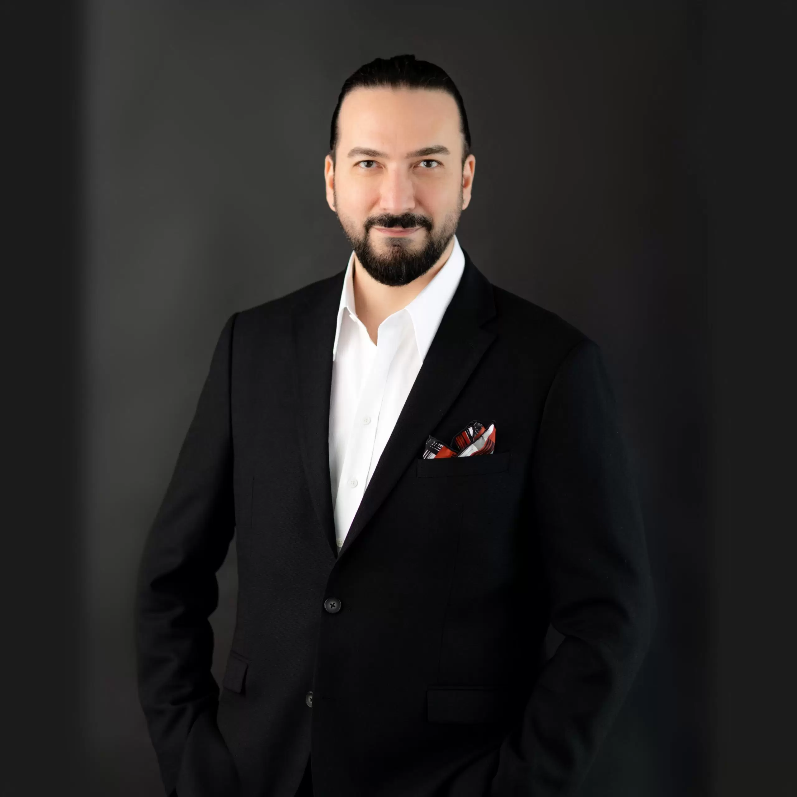
Prof. Dr. Murat Songu – Rhinoplasty (Nose Aesthetics) Specialist
Prof. Dr. Murat Songu was born in 1976 in Izmir and completed his medical education at the Ege University Faculty of Medicine. He then completed his residency training in the Department of Otorhinolaryngology at Celal Bayar University. Between 2005 and 2006, he received advanced training in rhinoplasty, functional nasal surgery, and skull base surgery in Bordeaux, France, working with leading rhinologists such as Prof. Vincent Darrouzet and Dr. Guy Lacher.
Prioritizing natural appearance, the preservation of breathing function, and facial aesthetic harmony, Prof. Dr. Songu is nationally and internationally recognized for his expertise in open rhinoplasty, piezo (ultrasonic) rhinoplasty, revision rhinoplasty, tipplasty, and functional septorhinoplasty. He has participated as an instructor and speaker at numerous rhinoplasty congresses worldwide.
With over 100 scientific publications, book chapters, and more than 1700 citations, Prof. Dr. Murat Songu is one of Türkiye’s most respected academics in the field of rhinoplasty, combining natural, facially harmonious, and functional results with scientific and aesthetic excellence.